Joints, or articulations, are such joints in which there is a gap or cavity between the articulating bones, filled with a lubricating fluid and surrounded by a connective tissue bag. The ends of the bones involved in the formation of the joint are covered with a thin layer of smooth articular cartilage, which facilitates the sliding of the bones. Consequently, the typical constituent elements of any joint are: the articular surfaces of the bones covered with cartilage, the articular bag and the articular cavity.
In the joint bag, it is necessary to distinguish between two layers: the outer, dense, playing a protective role, and the inner one, facing the cavity with its smooth surface, is the synovial layer. The latter has for the joint special meaning, as it releases a thick lubricating fluid that eliminates friction between the articular ends of the bones (synovial fluid).
In addition to the basic elements in some joints, there are additional devices. These include the articular lips, menisci, and discs. All these formations are found in those joints where the ends of the bones involved in their formation do not correspond to each other either in shape or in size of the articular areas.
Articular lips in the form of a narrow circular plate of cartilage are attached to the edges of a smaller bone, thereby increasing its surface. Articular discs are plates of cartilage located between articulating bones and fused at the edges with the articular bag. They divide the joint cavity into two isolated chambers. If there is a hole in the middle of the disc through which the chambers communicate with each other, such a disc is called a meniscus.
Ligaments should also be attributed to the additional adaptations of the joints. They are located either in the joint capsule itself, strengthening certain parts of the latter, or lie isolated at some distance from it, or, finally, are hidden inside the joints. In all cases, they act as brakes. The ligamentous apparatus regulates movements in a number of joints, limiting or completely stopping the mobility of bones in one direction and, conversely, allowing it in another.
The bones involved in the formation of joints are in full contact, in full contact with each other. The closure of the bone elements of the joint is determined by a number of factors, among which the musculature surrounding the joint is of primary importance. A certain degree of tension (tone), inherent in it even at rest, contributes to a tight connection with each other of the articular ends of the bones. The last factor that determines contact in the joints is the property of moist smooth articular
surfaces stick to each other, as well as the effect of atmospheric pressure. Movements in the joints are strictly natural. The nature of the movement is determined mainly by the shape of the articular areas of the articulating bones. In no other department of anatomy is the connection between form and function so clearly revealed as in the study of the joints. The shape of the articular areas of the bones can be compared with segments of geometric bodies of revolution. As you know, these bodies arise as a result of the rotation of a line (generator) around a straight fixed axis (axis of rotation). The shape of the bodies of revolution depends on the nature of the generatrix. If the latter is a straight line parallel to the axis of rotation, then the result is a cylinder. If such a generatrix is located at an angle to the axis of rotation, then a cone will be obtained.
In other cases, the generatrix may not be a straight line, but a broken line; then, as a result of the motion, we obtain other bodies of revolution. So, a semi-ellipse, rotating around an axis lying on its concave side, will give an ellipsoid of revolution, and a semicircle under the same conditions forms a ball.
The generatrix can be an arcuate curve, convexly facing the axis of rotation. In such cases, saddle-shaped surfaces of such bodies of revolution as a hyperboloid and others are obtained.
The study of the articular ends of different bones shows that their shape corresponds to the shape of the segments of the surfaces of a cylinder, a cone, a semi-ellipse, a ball, and a hyperboloid.
The nature of the movement of the bones in the joints corresponds to the movement of this "generating" around the fixed axis of rotation. Thus, one of the bones of the joint moves around another, immovable bone in a plane perpendicular to the axis of the given body of revolution. Consequently, the degree of mobility of one or another joint is determined mainly by the number of axes of movement in it. This sign is leading in the classification of joints.
There are uniaxial, biaxial and triaxial joints, as well as semi-movable and combined.
Uniaxial joints characterized by the fact that the geometric shape of the movements produced in them is due exclusively to the anatomical design of the joints; the difference in the work of the muscles is not reflected in the nature of the movements. The articular surfaces of the articulating bones correspond to each other in shape and are segments of geometric bodies of revolution formed around one axis. If the axis is located transversely, we get a block-shaped joint, if it is longitudinal, then it is cylindrical or rotatory.
Block joint has articular platforms resembling segments of a hyperboloid in shape. One of them, convex like a roller and having a groove in the middle, is called a block. The other, correspondingly concave, has a comb in the middle, which enters the furrow of the block. The axis of movement of the joint is frontal and is located transversely to the long axis of the articulating bones. The movements that take place in the block-shaped joint are in the nature of flexion and extension. The most typical example of block joints are the interphalangeal joints of the fingers.
In some block-like joints, the guide groove of the block lies not perpendicular to the axis of the latter, but at some angle to it. If continued, this furrow would form a helical line. Block-shaped joints of this type are called helical joints. An example is the shoulder joint.
Cylindrical (rotate) joint has articular platforms of a cylindrical or conical shape. Their axis coincides with the direction of the long axis of the articulating bones. The axis of movement of the joint runs vertically. Movements in a cylindrical joint are in the nature of rotation of the bone around its longitudinal axis to the outer and inner sides. An example of a rotator joint is the articulation between the radius and ulna.
Multiaxial joints characterized by articular surfaces of spherical shape. One of them forms a spherical head, the other - a correspondingly concave articular cavity.
Movements in spherical joints are performed around three main axes: around the frontal - flexion and extension; around the sagittal - abduction and adduction; around the vertical - rotation to the inner and outer sides. In addition to these movements around the main axes, others are possible, going along intermediate axes. This includes a circular motion, in which the end of the bone farthest from the joint describes a circle or oval, and the entire bone is a cone with its apex facing the center of the joint.
Spherical joints, in comparison with all others, are characterized by the greatest mobility, the range of motion in them is equal to the difference in the articular areas over their area. That is why, in the most mobile joints, the articular fossa is small compared to the size of the head (shoulder joint).
Strengthening of the spherical joints usually occurs due to some decrease in their mobility and is accomplished by increasing the contact surfaces of the bones. In such joints, the cavity is deeper and covers most of the head. The spherical joints are called the walnut joint. An example of a walnut joint is the hip joint.
Biaxial joints. There are two main types of biaxial joints - elliptical and saddle.
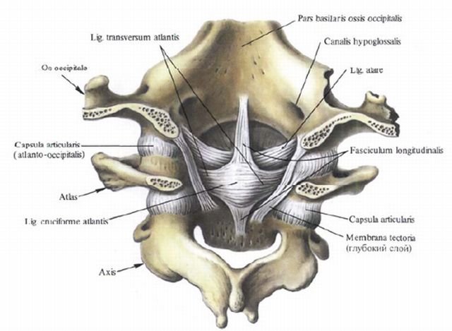
Elliptical joints have articular areas approaching in shape to a segment of the surface of an ellipsoid of revolution. Movements in elliptical joints are performed around two axes perpendicular to each other - frontal and sagittal. Around the first, flexion and extension are performed, around the second - abduction and adduction. An example of a typical elliptical joint is the wrist joint, as well as the atlantooccipital joint.
Saddle joints formed by two saddle-shaped articular surfaces placed on top of each other. In their geometric form, these surfaces resemble segments of an annular body of revolution. Movements in the saddle joints are made around two mutually perpendicular axes - frontal and sagittal, and one of the surfaces moves both along and across the other. The most typical saddle joint is the metacarpal joint. thumb brushes.
Semi-movable joints have almost flat articular surfaces, which are segments of the surfaces of bodies of revolution with a very large radius. Both articular areas are almost the same in their length, and therefore the movements in such joints are either completely absent or very insignificant.
The sacroiliac, intervertebral and some other joints are semi-movable.
Combined joints are a combination of several anatomically separate joints, acting as a whole. This group includes such joints that always work together, acting in the same direction. An example would be the superior and inferior radioulnar articulations (uniaxial), or both atlantooccipital articulations (biaxial).
There are more complex built combined joints. These are most often two consecutive and anatomically separate joints, separated by either one or several bones connected into a single whole. The most typical joints of this type are the combined joints of the hand and foot.
Thus, the combined joint is not an anatomical, but a physiological concept.
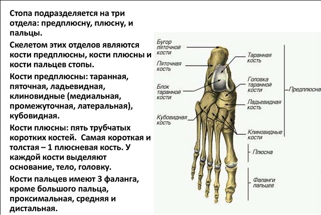
Marked changes in the shape and structure of bones under the influence of physical activity may also concern the structure and shape of whole parts of the body. It is known that between the human foot and hand, on the one hand, there is a significant similarity, on the other hand, there are significant differences (Fig. 23). The similarity is explained by the fact that in the distant ancestors of man, the upper and lower limbs performed approximately the same functions - they served for movement. When a person switched to upright walking, the upper limbs gradually turned from organs of support and movement into labor organs. The functions of the hand began to differ sharply from the functions of the foot, and the shape changed accordingly. Examples modern life give us amazing confirmation of this law. Some people, deprived of upper limbs from birth or for other reasons, are taught from early childhood to do with the foot the work that is usually done with the hand.
Rice. 23. Skeleton of the hand (a) and foot (b) of a person.
1 - bones of the wrist; 2 - bones of the metacarpus; 3 - phalanges of fingers.
And it should be noted that such a foot is adapted to perform very delicate work. An example of this is the activity of the artist Untan, who was born without arms, and some carpet embroiderers who were deprived of upper limbs.
In many ways, the structure of bones is determined by nutrition. The intake of mineral salts and vitamins D, A and C is of paramount importance. Without vitamin D in bone tissue salts cannot be deposited and rickets develops. With a lack of vitamin A, a violation of the activity of bone cells occurs, resulting in the formation of abnormal thickening of the bones and a decrease in bone cavities and channels. Bone growth and chemical composition regulated by the endocrine glands and the nervous system.

Rice. 24. Scheme of the structure of the joint in the context:
1 - joint cavity;
2 - articular surfaces covered with cartilage;
3 - fibrous layer of the articular bag;
4 - synovial layer of the articular bag.
The bones are interconnected by ligaments, cartilage and joints, which have a complex structure (Fig. 24). The joint is formed by special surfaces of bones covered with cartilage. It is surrounded by a capsule that hermetically separates its cavity from the surrounding tissues. The capsule of the joint and the bones that form it are strengthened by ligaments. The inner surface of the joint capsule (the so-called synovial layer) secretes a special synovial fluid that moisturizes the articular surfaces of the bones and thus acts as a lubricant that reduces friction between the bones. There is no air in the cavity of the joints, therefore, there is a negative pressure, therefore, the outside air exerts pressure on the articular surfaces of the bones adjacent to each other and presses them against each other the stronger, the larger the joint. The joints of the bones are very strong and at the same time provide a high degree of mobility and flexibility of the body. Many circus performers, acrobats and athletes amaze us with such extraordinary flexibility that it seems as if their body is “without bones”. This development of joint mobility is achieved by special exercises, persistently and systematically carried out from early childhood, when the flexibility of the joints is much greater than in adulthood.
1125 0
Perfect glide for mindless movement
When you see another “snake woman” in the “Minute of Glory”, twisting her body almost into pigtails, you understand that the structure of joints and bones that is standard for other people is not about her. What kind of dense fabrics can we talk about - they simply do not exist here!
However, even her hard tissues have a place to be - a lot of joints, bones, as well as structures for their connections, according to the classification, divided into several categories.
Bone classification
There are several types of bones depending on their shape.
Tubular bones with a bone marrow cavity inside and formed from compact and spongy substances, performing supporting, protective and motor roles. Subdivided into:
- long(bones of the shoulders, forearms, hips, legs), which have a bi-epiphyseal nature of ossification;
- short(bones of both wrists, metatarsals, digital phalanges) with a monoepiphyseal type of ossification.

Bones of a spongy structure, with a predominance of a spongy substance in the mass with a small thickness of the covering layer of a compact substance. Also divided into:
- long(including costal and sternal);
- short(vertebral, carpal, tarsal bones).
This category also includes sesamoid bone formations, located near the joints, participating in their strengthening and contributing to their activity, having no close connection with the skeleton.
Flat shaped bones, including categories:
- flat cranial(frontal and parietal), performing the role of protection and formed from two outer plates of a compact substance with a layer of spongy substance located between them, having a connective tissue genesis;
- flat bones of both limb girdles(scapular and pelvic) with a predominance in the structure of a spongy substance, acting as a support and protection, with a genesis from cartilaginous tissue.
Bones of mixed (endesmal and endochondral) genesis with different structure and tasks:
- forming the base of the skull;
- clavicular.
Only the bones do not live on their own - they are interconnected by joints in the most ingenious ways: two, three, at different angles, with varying degrees of sliding over each other. Thanks to this, our body is provided with incredible freedom of static and dynamic postures.

Synarthrosis VS diarthrosis
But not all bone joints should be considered diarthrosis.
According to the classification of bone joints, the following types of articulation do not belong to these:
- continuous (also called adhesions, or synarthrosis);
- semi-movable.
The first grade is:
- synostoses- fusion of the boundaries of the bones between themselves to complete immobility, zigzag "lightning" of the seams in the cranial vault;
- synchondrosis- fusion by means of a cartilaginous layer, for example, an intervertebral disc;
- syndesmoses- strong "stitching" of the connective tissue structure, the interosseous sacroiliac ligament, for example;
- synsarcoses- when connecting bones with the help of a muscular layer.
The tendon membranes stretched between the paired formations of the forearms and lower legs, holding them dead next to each other, are also not joints.

As well as semi-mobile joints (hemiarthrosis) in the face of the pubic symphysis with a small (incomplete) cavity-gap in the thickness of the fibrocartilaginous suture, or in the form of sacroiliac amphiarthrosis with real articular surfaces, but with an extremely limited range of motion in the semi-joints.
Structure and functions
A joint (discontinuous or synovial connection) can only be considered a movable joint of bones that has all the necessary attributes.
In order for all dysarthrosis to move, there are special formations and auxiliary elements in them in strictly defined places.

Structure diagram knee joint
If on one bone it is a head, which has a pronounced roundness in the form of a thickening - the epiphysis of the end section, then on the other associated with it, it is a depression exactly corresponding to it in size and shape, sometimes significant (such in the pelvic bone for its vastness is called "vinegar"). But there may also be an articulation of one bone head with a structure on the body-diaphysis of another, as is the case in the radioulnar joint.
In addition to the ideal correspondence between the forms that form the joint, their surfaces are covered with a thick layer of hyaline cartilage with literally mirror-smooth surface for flawless sliding over each other.
But smoothness alone is not enough - the joint should not crumble into its component parts. Therefore, it is surrounded by a dense elastic connective tissue cuff - a capsule bag, similar to a lady's muff for warming hands in winter. In addition, its fastening is served by a ligamentous apparatus of different power and muscle tone, which ensures biodynamic balance in the system.
A sign of true dysarthrosis is the presence of a full-fledged joint cavity filled with synovial fluid produced by cartilage cells.
The classic and simplest in structure is the shoulder. This is a joint gap between its bag and two bone endings that have surfaces: the round head of the humerus and the articular cavity on the scapula that matches in configuration, filled with synovial fluid, plus ligaments that hold the entire structure together.
Other dysarthroses have a more complex structure - in the wrist, each bone contacts several adjacent ones at once.
The spine as a special case
But the relationships between the vertebrae are especially complex - short-columnar bones that have a complex surface topography and many structures for varying degrees of movable adhesion with neighboring formations.
The spine has a structure resembling a rosary, only its "beads" are the bodies of each of the neighboring bones, which are interconnected by means of hemiarthrosis (synchondrosis) based on a cartilaginous disk. Their spinous processes, which overlap each other like tiles, and the arches, which form a receptacle for the spinal cord, are fastened with rigid ligaments.
The joints between the transverse processes of the vertebrae with flat surfaces (as well as the costovertebral joints, formed by means of the costal heads and articular cavities on the bodies of the vertebrae located laterally) are quite real, having all the necessary attributes: working surfaces, cracks, capsules and ligaments.
In addition to connections with each other and with the ribs, the vertebrae form an fusion in the area of the sacrum, which turns this group into a monolith, to which, through real joints, a “tail”-coccyx is attached - the formation is quite mobile, especially during childbirth.
Dysarthroses are the beginning of the pelvic girdle, formed by the bones of the same name, in front in the center closing in a ring by the pubic symphysis.
In addition to the intervertebral joints, there are other joints in the support column system: a combination that forms one unpaired and two paired components of the atlanto-axial connection (between the I and II vertebrae) and paired atlanto-occipital (between the I vertebra and the occipital bone).
Due to this very structure, the spine is an incredibly flexible formation, having a large degree of freedom of movement and at the same time exceptionally strong, bearing the entire weight of the body. In addition to the support function, it also performs a protective role, serving as a channel through which the spinal cord passes, and is involved in hematopoiesis.
The spectrum of damage to the joints of the vertebrae is diverse: from injuries (with various categories and displacements) to metabolic-dystrophic processes leading to varying degrees of stiffness of the spine (and similar conditions), as well as infectious lesions (in the form of them, lues, brucellosis).
Detailed classification
The above classification of bone joints does not include the taxonomy of the joints, which has several options.
In accordance with the number of articular surfaces, the following categories are distinguished:
- simple, with two surfaces, as in the joint between the phalanges of the first finger;
- complex in the presence of more than two surfaces, for example, in the elbow;
- complex with the presence of internal cartilaginous structures dividing the cavity into non-isolated chambers, as in the knee;
- combined as a combination of joints isolated from each other: in the temporomandibular joint, the intraarticular disk divides the working cavity into two separate chambers.
According to the functions performed, joints with one, two and multiple axes of rotation (one-, two- and multi-axis) are distinguished, depending on the shape, having the form:

An example of uniaxial joints are:
- cylindrical - atlanto-axial median;
- block-shaped - interphalangeal;
- helical - shoulder-elbow.
Structures of complex shape:
- ellipsoid, like a radiocarpal lateral;
- condylar, like a knee;
- saddle-shaped, like the metacarpal-carpal joint of the first finger.
Multiaxial are represented by varieties:
- spherical, like a shoulder;
- cup-shaped - a deeper variation of the spherical (like the hip);
- flat (like intervertebral).

Radioulnar cylindrical joint
There is also a separate category of tight joints (amphiarthrosis), which differ in the shape of the surfaces, but are similar in another - they are extremely stiff due to the strong tension of the capsules and a very powerful ligamentous apparatus, therefore their sliding displacement relative to each other is almost imperceptible.
Characteristics, design and functions of the main joints
With all the abundance of joints in the human skeleton, it is most logical to consider them as separate groups - categories of joints:
- skulls;
- spine;
- limb belts (upper and lower).
cranial joints
In accordance with this provision, two diarthroses enter the skeleton of the skull:
- temporomandibular;
- atlanto-occipital.
The first of these paired connections was created with the participation of the heads of the bone mandible and working depressions on the temporal bones.
The joint consists of two synchronously functioning, although spaced apart on opposite sides of the skull formations. It is condylar in configuration, belongs to the category of combined ones due to the presence in it of a cartilaginous disk dividing its volume into two chambers isolated from each other.
Due to the existence of this diarthrosis, the freedom of movement of the lower jaw in three planes and its participation both in the process of primary food processing and in swallowing, breathing and the formation of speech sounds are possible. The jaw also serves as a means of protecting the organs of the oral cavity from damage and is involved in creating the relief of the face. It can be subjected to both injury and infection during the development of acute (mumps) and exacerbation of chronic (tuberculosis,) diseases.
The configuration of the paired atlanto-occipital region is also condylar. It serves to connect the skull (its occipital bone with convex working surfaces) with the spine through the first two cervical vertebrae, acting as a single unit, on the first of which - the atlas - there are working fossae. Each half of this synchronously functioning formation has its own capsule.
Being a biaxial atlas, it allows head movements both according to the frontal and sagittal axes - both nodding and tilting left and right, providing freedom of orientation and the fulfillment of a social role by a person.
The main pathology of the atlanto-occipital diarthrosis is trauma as a result of a sharp tilting of the head and the development of osteochondrosis and other metabolic-dystrophic conditions due to the long-term maintenance of a forced posture.
Shoulder girdle
Given the above description of the spine, turning to diarthrosis of the shoulder girdle, it should be understood that the joints  clavicle with sternum and scapula with clavicle are synarthroses. The real joints are:
- brachial;
- elbow;
- radiocarpal;
- carpal-metacarpal;
- metacarpophalangeal;
- interphalangeal.
The sphericity of the head of the humerus is the key to almost complete circular freedom of rotation of the upper limb, therefore, the shoulder refers to multiaxial joints. The second component of the mechanism is the scapular cavity. All other attributes of diarthrosis are also present here. The shoulder connection is most susceptible to damage (due to the large degree of freedom), to a much lesser extent - to infections.

The shoulder joint is the most mobile in the entire ODA
The complex structure of the elbow is due to the articulation of three bones at once: the humerus, radius and ulna, which have a common capsule.
The shoulder-elbow joint is trochlear: the shoulder block enters the notch on the ulna, the shoulder-radius joint is the result of the head of the condyle of the shoulder entering the fossa of the head of the bone-ray with the formation of a spherical working area.
Movements in the system are carried out according to two axes: flexion-extension, and also due to the participation of the proximal radioulnar joint, rotation (pronation and supination) is possible, because the head of the beam rolls along the groove on the ulna.
The problems of the elbow joint are damage, as well as inflammatory conditions (with acute and exacerbation of chronic infections), dystrophy due to professional sports.
The radioulnar distal joint is a cylindrical joint that provides vertical rotation of the forearm. In the working cavity, there is a disc that separates the designated joint from the cavity of the carpal joint.
Diseases of the elbow area:
- instability;
- stiffness.
 By means of a capsule covering the lower epiphysis of the beam and the first row of carpal bones, an elliptical configuration of the wrist joint is formed. This is a complex articulation with sagittal and frontal axes of rotation, allowing both adduction-abduction of the hand with its circular rotation, and extension-flexion.
The most common diseases:
- injuries (in the form of bruises, fractures, sprains, dislocations);
- synovitis;
- varying degrees of severity of carpal tunnel syndrome;
- arthritis and hip;
- knee;
- ankle;
- tarsal-metatarsal;
- metatarsophalangeal;
- interphalangeal.
The shape of the hip multiaxial joint is bowl-shaped, with the participation of the head femur and the ischial cavity, which provides adduction-abduction of the thigh forward-backward and medially-laterally, as well as its rotation.
TSB is susceptible to damage (due to the high degree of freedom) and damage by microbial flora, most often brought here hematogenously (tuberculosis, brucellosis, gonorrhea).
The most common diseases of the hip area: 
- bursitis;
- tendinitis;
- femoral-acetabular impingement syndrome; .
- extension-flexion;
- slight vertical abduction-adduction (in flexion position).
- rammed;
- ram-heel-navicular;
- calcaneocuboid;
- sphenoid-navicular.
The structure of diarthrosis allows you to:
The most common disorder of function - (external or internal), as well as a violation metabolic processes in the body and blood circulation in the lower extremities.
The tarsal area is formed by a "mosaic" of the joints:
These are compounds of a combined or flat configuration (the first two are cylindrical and spherical).
Tarsal-metatarsal diarthroses are represented by various (mostly flat) joints that form a support for the arches of the foot, made by metatarsophalangeal (block-shaped) joints.
Also, the block-shaped interphalangeal joints of the feet give the toes a sufficient level of mobility and flexibility (patients who have lost both hands draw and even sew with their feet) without sacrificing strength.
Small joints of the feet tend to be damaged due to metabolic and dystrophic processes in the body, with disorders of local and general blood supply and as a result of chronic injuries in the form of wearing shoes with high heels or simply tight.
Existence various ways bone joints, as well as the diversity of the articular surfaces themselves, understanding their structure and function allows a person not only to live and act, but also to treat the musculoskeletal system (and, if necessary, even replace structures that have become unusable with artificial ones).
The most common, most mobile connections in which precise dosed movements are made in certain directions. Mandatory conditions for the formation of a joint: 1) articular surfaces; 2) the presence of an articular bag; 3) articular cavity; 4) synovial fluid.
1. Articular surfaces bones are covered with cartilage, which gives the bones smoothness, contributing to their better glide, and elasticity, softening the shocks during movements. Articular surfaces that correspond to each other are called congruent. Their shape is compared to geometric shapes: as surfaces resulting from the rotation of a straight line or a curve around a conditional line (I- block-shaped; II-ellipsoid; III- saddle; IV - spherical; A - bones of the hand; one- distal phalanx; 2-middle phalanx; 3- head of the phalanx; 4- phalanges (finger bones); 5 - proximal phalanx; 6- base of the phalanx; 7- body of the phalanx; 8- head of the metacarpal bone; 9 - third metacarpal bone; 10 - body of the metacarpal bone; 11 - base of the metacarpal bone; 12- metacarpus (I-V metacarpal bones); 13 - styloid process; 14 - trapezoid bone; 15- trapezoid bone; 16 - capitate bone; 17- hook-shaped bone; 18- trihedral bone; 19 - pisiform bone; 20 - lunate bone; 21 - navicular bone.). For example, when a curved line is rotated, a sphere, an ellipse, or a block can be formed; when a straight line is rotated, a cylinder is obtained.
* AT cylindrical joint only rotation takes place.
* AT trochlear joints articular surface in the form of a transversely lying cylinder, the long axis of which lies transversely, in the frontal plane, perpendicular to the long axis of the articulating bones. In them, only movements around one, frontal, axis are possible (flexion and extension).
*Elliptical joint. Two surfaces in the form of an ellipse. One of them is convex and the other is concave. Movements are possible around two mutually perpendicular axes. For example, the wrist joint - flexion and extension around the frontal axis, adduction and abduction - around the sagittal axis
* If the joint has an articular head in the form of a protruding elliptical process, then the difference in size and shape between the articulating surfaces is large, and the joints are also called condylar. Movement in the condylar joint occurs around the frontal axis and around the longitudinal (rotation). For example, when the head moves, the 1st cervical vertebra in the form of a ring rotates around the odontoid process of the 2nd vertebra.
* AT saddle joint 2 surfaces sit on top of each other, one of them moves along and across the other. It can move around two mutually perpendicular axes. For example, in the carpometacarpal joint of the thumb, not only abduction and adduction takes place, but also opposition of the thumb to the rest.
* ball joint. Often one articular surface is shaped like a head and the other is shaped like a cavity. Movement occurs around three axes, and when moving from one axis to another, a circular motion is obtained. The greater the difference between the length of the arc of the head and the arc of the cavity, the greater the range of motion.
* AT flat joint the articular surfaces of the bones are flat and slide relative to each other during movements. Flat joints (wrists, tarsals) are stiff, but movements are possible around three axes.
2. Articular bag (geometric bag) has 2 shells: outer - fibrous (strong), fused with the periosteum, and synovial - inner - with the edges of the articular cartilage. synovial membrane covered with a layer of endothelial cells, has a smooth shiny appearance, forms villi that increase its surface. This shell produces synovial fluid, which moisturizes the articular surfaces, eliminating friction of the bones, and absorbs fluid, ensuring metabolism. Synovial fluid is formed from blood plasma, consists of hyaluronic acid and tissue fluid, is similar in appearance to egg white. In sedentary or immobile joints, the synovial fluid becomes viscous, but if the joints begin to move actively, the viscosity decreases. Joint fluid contains phagocytes that destroy microorganisms and substances that enter the joint when it is damaged. In some places, the joint capsule becomes thinner and a protrusion forms. synovial bursa. Such bags are located under the muscles or tendons and reduce their friction against the bone during movement.
Let's try to understand this complex mechanism, where each bone occupies certain place and is in direct connection with one or more neighboring bones. The exceptions are the so-called sesamoid bones, located in the thickness of the tendons of the muscles (for example, the patella and pisiform bone of the wrist), and the hyoid bone. The mobility of body parts depends on the nature of the joints between the bones.
There are continuous connections that form strong fixed or inactive structures, discontinuous connections, or joints that allow the bones to move relative to each other, as well as a transitional type of connections - semi-joints, or symphyses.
Connective tissues
In continuous joints, the bones are interconnected by a layer of connective tissue, devoid of any gaps or cavities. Depending on the type of connective tissue, fibrous, cartilaginous and bone continuous connections are distinguished.
Fibrous connections include numerous ligaments, interosseous membranes, sutures between the bones of the skull, and connections between teeth and jaws (Fig. 1). Ligaments are dense bundles of fibers that run from one bone to another. There are a lot of ligaments in the region of the spine: they are located between individual vertebrae; during movements of the spinal column, they limit excessive inclinations and contribute to the return to the starting position. The loss of elastic properties by these ligaments in old age can lead to the formation of a hump.
Interosseous membranes have the form of plates stretched between the bones for a considerable length. They firmly hold one bone near another, serve as a place of attachment of muscles. Such membranes are located, for example, between the long tubular bones of the forearm and lower leg.
Skull sutures
The sutures of the skull are the connections between the bones of the skull with the help of thin layers of fibrous connective tissue. Depending on the shape of the edges of the skull bones, serrated, scaly and flat sutures are distinguished. The most elegant flat suture is found only in the region of the facial region of the skull, and a strong jagged suture, similar to a zipper, is found in the roof of the brain region. The temporal bone, like fish scales (hence the name of the suture), is fixed on the lateral surface of the skull.
spring
In a newborn child, there are no sutures, and significant membranous spaces between the bones of the skull are called fontanelles. Due to the presence of fontanelles, the shape of the skull can change during the passage of the fetus through the birth canal, which facilitates the birth of a child. The largest anterior, or frontal, fontanel is located in the region of the crown, has a diamond shape and disappears only in the second year of life. Smaller fontanelles, located in the occipital and temporal regions of the skull, close on the 2-3rd month after birth. The formation of seams ends by 3-5 years of age. After 30 years, the seams between the bones of the skull begin to overgrow (ossify), which is associated with the deposition of calcium salts in them. In men, this process occurs somewhat earlier than in women. In old age, the human skull becomes smooth, the boundaries between the bones are virtually indistinguishable.
Teeth
The teeth are fixed in the cells (alveoli) of the jaws with the help of the so-called periodontium - bundles of strong fibers that connect the root of the tooth with the surface of the alveoli. Experts call this type of connection “impacting”, however, paying attention to some anatomical discrepancy: after all, the teeth grow from the inside of the jaw, and are not driven into it from the outside!
Intervertebral discs
Continuous connections of bones with the help of cartilaginous tissue are distinguished by strength, elasticity and low mobility, the degree of which depends on the thickness of the cartilage layer. This type of connection includes, for example, intervertebral discs (see Fig. 1), the thickness of which in the lumbar, most mobile, section of the spinal column reaches 10-12 mm. In the center of the disc is an elastic nucleus pulposus, which is surrounded by a strong fibrous ring. The core is strongly compressed and constantly strives to expand, therefore it springs and absorbs shocks like a buffer. With excessive loads and injuries, the intervertebral discs can be deformed, displaced, as a result, the mobility and depreciation properties of the spine are impaired. With age, in case of metabolic disorders, calcification of the intervertebral discs and ligaments, the formation of bone growths on the vertebrae can occur. This process, called osteochondrosis, also leads to limited mobility of the spinal column.
Continuous cartilage connections
Many continuous cartilaginous connections between bones are present only in childhood. With age, they ossify and turn into continuous bone joints. An example is the fusion of the sacral vertebrae into a single bone - the sacrum, which occurs at the age of 17-25. The formation of some bones of the skull (for example, the occipital, temporal) from several separate parts is observed at the age of 1 to 6 years. Finally, the fusion of ends tubular bones with their middle part in the period from 17 to 21 years for women and from 19 to 23 years for men, it determines the completion of growth processes.
Joints and semi-joints

Semi-joints are also cartilaginous connections between bones. But in this case, in the thickness of the cartilage there is a small slit-like cavity filled with liquid, which increases the mobility of the joint. The semi-joint is the pubic symphysis - the connection of two pelvic bones to each other in front. The possibility of a slight divergence of the pelvic bones in the symphysis area is important for women in the process of childbirth.
Joints are movable joints between bones. They are discontinuous joints that always have a slit-like space between the connecting bones. In addition to the slit-like articular cavity in each joint, the articular surfaces of the articulating bones and the articular capsule surrounding it from all sides are distinguished (Fig. 2).
Articular capsule and articular cartilage
The articular surfaces of the articulating bones are covered with a layer of smooth articular cartilage 0.2 to 6 mm thick, which reduces friction between moving bones. The greater the load, the thicker the articular cartilage. Since the cartilage has no vessels, the main role in its nutrition is played by the synovial fluid that fills the joint cavity.
synovial membrane
The articular capsule surrounds the articular cavity and adheres to the bones along the edge of their articular surfaces or slightly away from it. The joint capsule consists of two layers: the outer one is a dense fibrous membrane and the inner one is a thin synovial membrane. It is the synovial membrane that secretes a transparent, viscous synovial fluid into the joint cavity - a kind of lubricant that facilitates the sliding of the articulating bones. The synovial membrane can form various outgrowths: folds inside the joint, which serve to cushion during movement, as well as protrusions outside the joint capsule, called bags (bursae). Being located around the joint in the form of soft pads under the tendons of the muscles, the bags reduce the friction of the tendons on the bone during movements in the joint. As a result of bruises, inflammation of the bag can develop - bursitis. In this case, the bags (and the joint area) swell due to an increase in the volume of the fluid filling them.
Discs and menisci
The joint cavity has a slit-like shape due to the tight contact of the articular cartilage and the negative pressure inside the joint. To increase the similarity of the contacting surfaces, additional cartilage pads can be placed in the joint cavity: discs and menisci (crescent-shaped plates). They perform a shock-absorbing function and contribute to a variety of movements in the joint. For example, in the knee joint there are two menisci, and in the joints of the lower jaw there are discs.
Bundles
Retention of the bones in the articular state is facilitated by contractions of the muscles surrounding the joint. This is also served by ligaments that can be located in the joint cavity (as, for example, strong cruciate ligaments of the knee joint) or on top of its capsule. Ligaments strengthen the joint capsule, direct and limit movement. As a result of trauma, an unsuccessful movement, stretching and even rupture of the ligaments can occur, resulting in a displacement of the bones in the joint - dislocation.
Simple and complex joints

If two bones are connected in a joint, then it is called a simple joint. In complex joints, several bones are articulated (for example, in the elbow - three bones). In cases where movements in two independent joints occur simultaneously (right and left joints of the lower jaw), they speak of a combined joint.
To characterize movements in the joints, three conditional mutually perpendicular axes are used, around which movements are made. According to the number of axes, multiaxial joints are distinguished, in which movements occur around all three axes of three-dimensional space, as well as biaxial and uniaxial joints. The nature and scope of movements in the joint depend on the features of its structure, primarily on the shape of the articular surfaces of the bones. The relief of the articular surfaces is compared with geometric bodies, therefore, spherical (multiaxial), elliptical (biaxial), cylindrical and block-shaped (uniaxial), flat and other joints are distinguished (Fig. 3).
One of the most mobile is the spherical shoulder joint (Fig. 4), in which the round head of the humerus articulates with the glenoid cavity of the scapula. Arm movements in the shoulder joint are possible around all axes. In flat joints (for example, between the sacrum and pelvic bones), mobility, on the contrary, is extremely small.
muscles
Joints are formed under the influence of muscle activity, and their structure is closely related to function. This law operates both in the process of evolution and during the individual development of the organism. An example is the features of the skeleton of the upper and lower limbs of a person, which in both cases has a general structural plan, but differs in the fine organization of bones and their joints.
In the skeleton of the limbs, a belt is distinguished (shoulder and pelvic) and a free limb, which includes three parts: shoulder, forearm and hand upper limb; thigh, lower leg and foot at the bottom. Differences in the structure of the skeleton of the limbs are due to their different functions. The upper limb is an organ of labor adapted to perform various and precise movements. Therefore, the bones of the upper limb are relatively smaller and are connected to each other and to the body by very mobile joints. The lower limb in humans is designed to support the body and move it in space. The bones of the lower limb are massive, strong, and the joints have dense capsules, a powerful ligamentous apparatus, which limits the range of motion.
Hand and foot

The main differences are observed in the structure of the hand and foot. There are many movable joints among the joints of the hand, as a result of which various subtle movements can be carried out. The joints of the thumb are especially important, due to which it is possible to oppose the thumb to all others, which contributes to the capture of objects. The joints of the hand reach such development only in humans! The foot bears all the weight human body. Due to the vaulted structure, it has spring properties. Flattening of the arches of the foot (flat feet) leads to rapid fatigue when walking.
Joint mobility increases under the influence of training - remember the amazing agility of athletes and circus acrobats. But even ordinary people need to move more in order to maintain good joint mobility. In children, the joints are usually more mobile than in adults and especially the elderly. This is due to a decrease in the elasticity of the ligamentous apparatus with age, abrasion of articular cartilage and other reasons.
Head healer - movement
Limitation of mobility and pain during movements in the joint may be associated with the gradual destruction of articular cartilage and impaired production of synovial fluid. At the same time, the articular cartilage gradually becomes thinner, cracks, the amount of lubrication becomes insufficient - as a result, the range of motion in the joint decreases. To prevent this from happening, you should conduct a mobile healthy lifestyle life, eat right, and, if necessary, strictly follow the doctor's instructions, because life is movement, and movement is impossible without a clear work of the musculoskeletal system.